Keratoma
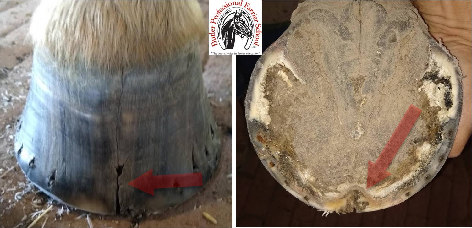
Keratoma is identified by a persistent, vertical indentation or crack in the dorsal (front of the) hoof wall (right) and an inward deviation of the white line towards the sole (left). This horse was not in pain from the keratoma and was able to perform its functions as a trail horse as long as it wore a shoe.
When it comes to diseases of the horse’s foot, keratoma is not as scary as it sounds. Literally, keratoma means a tumor (-oma) in the hoof wall (keratin). Tumor can sound intimidating, but most of the time these growths are benign and don’t cause pain. In instances where the horse is sound and does not appear to be in pain, usually the keratoma is left alone and all that is left is a blemish in the hoof wall. Keratomas are rare.
Keratoma occurs between the inner hoof wall and bone, usually at the toe. In cases where the horse is in pain, the most common cause of the pain is the tumor is pressing on the sensitive laminae and the coffin bone.
In some cases, Keratoma may be noticeable. There may be swelling in the coronary band, a persistent, vertical indentation or crack down the front of the hoof wall, and the white line may appear bent in toward the center of the sole.
In a case I worked on this summer, the vertical crack down the front of the hoof was so deep and persistent that the horse appeared to have a cloven hoof like a cow or deer. Strangely, the horse did not appear to be in pain and we were able to treat the condition by simply applying a shoe to stabilize hoof.
There are a few possible causes for a Keratoma to develop in the horse’s foot. As is the case in many other diseases of the foot, genetics is can always be a factor. Besides genetic predisposition to develop keratoma, trauma to the coronary band and persistent sand cracks may be the culprit in the development of the tumor.
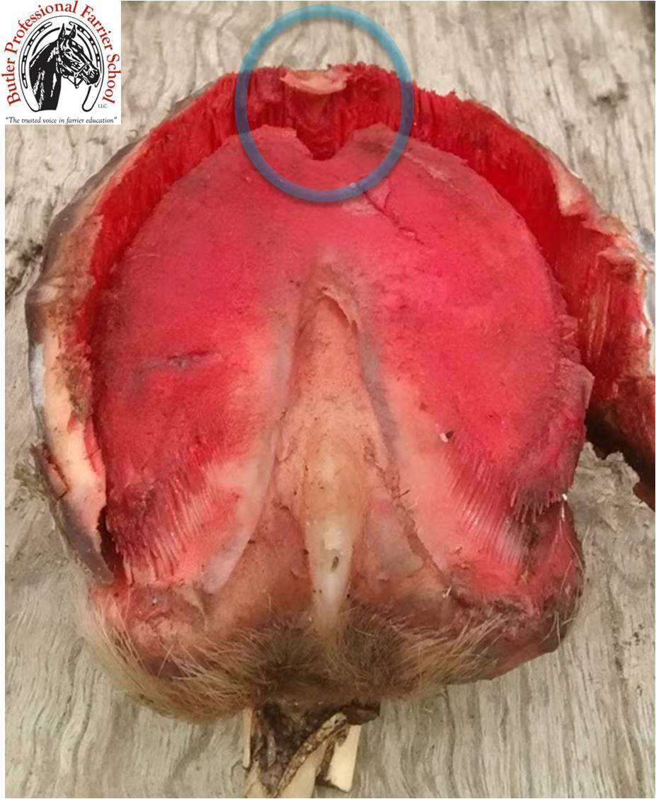
This cadaver specimen foot was used in a dissection to show the effect of a persistent keratoma on the coffin bone (circle). In cases, where this tumor affects the horse’s comfort, the veterinarian may remove the keratoma surgically.
Treatment for Keratoma varies. Like the example, I mentioned before, simply applying a shoe (sometimes with side clips) can help to stabilize the hoof capsule. In severe cases, where pain is present, the veterinarian should be called upon to surgically remove the keratoma. Farriers should not do this! The veterinarian does the surgery while the horse is under general anesthesia. Usually, the vet will use a motorized burr to quickly remove diseased hoof wall. The farrier should be present to apply a side clipped shoe to hold the foot together before surgery. It is easier to apply the shoe before the surgery has taken place so the farrier doesn’t have to work around the operated-on area. The shoe should also be applied so that the affected part of the foot does not touch the shoe. A hospital plate may also be useful to prevent infection in the bottom of the hoof. These are rare cases.
One time, a concerned horse owner brought me pictures of his horse with the noticeable inwardly deviated white line on the bottom of the foot. There appeared to be no indentation on the dorsal hoof wall. I asked if the horse was lame and how the horse’s feet were being cared for presently. He replied that the horse appeared to be very sound, the hoof appeared otherwise healthy, and that they were simply trimming the horse. In this case, continuing the hoof maintenance plan of trimming the horse and watching for any further development is all that was needed. There is wisdom in the adage, “If it isn’t broke, don’t fix it!”
Related Posts
-
Horses are living animals so they will inevitably move aroun...
-
©2013 Doug Butler PhD, CJF, FWCF Butler Professional Farri...
-
Each year around Valentine’s Day, we are reminded to t...