Osteomyelitis
Osteomyelitis is a painful infection of the coffin bone (P3) that can follow laminitis/founder. Prevention is the best treatment.
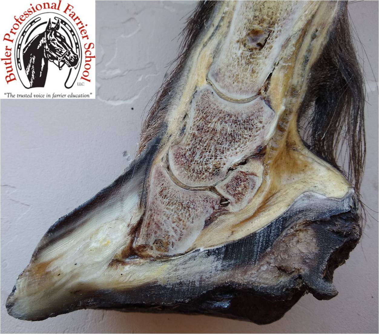
Osteomyelitis is distinguished from pedal osteitis in that the former implies infection of bone; the later implies inflammation due to bruising of the coffin bone. Bone pain is some of the most severe hurt an animal can experience. Osteomyelitis can be a sequela (following the original disease) to severe founder that results in sole perforation or from neglected foot abscesses. It also may result from a sequestrum (piece of dead or necrotic bone) that may result from a coffin bone fracture or a puncture wound in the sole.
Usually, only the outer border of the coffin bone or distal phalanx is affected in founder cases. The blood circulation coming from the circumflex artery that supplies the bone with nourishment is interrupted. The ischemia (lack of blood supply) caused by the destruction of this artery also affects the sensitive sole since most of its nourishment comes from this source. Necrosis (tissue death) may spread to other bones. The infection becomes almost impossible to stop once it spreads due to the difficulty of getting antibiotics to the infection site because of the compromised circulation.
Afflicted horses will exhibit signs of extreme bone pain. They will place their legs underneath the body and try to press downward by standing on their toes. Sole abscesses are usually present. In some cases the horse will appear to have a superficial flexor contracture with the fetlock knuckled over. Horses often develop dicubital ulcers (bed sores) on boney prominences from lying down. Radiographs will reveal areas of osteomiosis (disintegration of bone) in the coffin bone. White blood cell count is usually normal. However, a bone scraping biopsy is usually positive for Staphlococcus bacteria.
George Platt DVM and Burney Chapman CJF found medical treatments with antibiotics to be of little value in more than a hundred cases they treated due to the lack of circulation in the infected areas. In those cases where they did have success, Dr. Platt removed soft necrotic(dead) bone through the dorsal hoof wall and the sole surgically with a curet (bone scraping instrument). Mr. Chapman applied a heartbar shoe with a treatment plate and packed the wound with sugardine (a syrupy mixture of betadine and table sugar) and changed the bandage every other day until granulation tissue covered the bone. Dr. Platt recommended administering the antibiotic Baytril 5 mg/kg and Eq Stim (made by Neogen Corp.) 1cc/250 pds on day 1 and day 4. He repeated the same dose every week. They both concluded that once established, the bone infection was almost impossible to cure. The prognosis (expected outcome) was grave. Prevention is the best cure.
Prevention is best achieved with a treatment plate when sole abscesses are present and/or the coffin bone is severely foundered. This holds medication in place and prevents pressure and contamination of the wound. The plate is bolted to the ground surface of the shoe with 5/16th inch bolts and can be made from 1/8 inch aluminum or ¼ inch thick copolymer plastic. We prefer the plastic plate since it is 1) less likely to deform, 2) does not have sharp edges and 3) the bolts stay in place without lock washers. We recommend the antiseptic and poultice packing be changed frequently by the owner.
Related Posts
-
Last week, we talked about getting horses to stand still. We...
-
Farriers are presented with different horse feet scen...
-
Last week, I interviewed and wrote about Martha Crawford Can...